![]()
![]()
Journal of Cranio-Maxillofacial Surgery
Volume 38, Issue 3,
April 2010, Pages 160-165
|
|
|
|
|
Osteogenic uni- or bilateral form of the guided rapid maxillary expansion
K. Al-Ouf1,
![]() ,
,
![]() ,
Head of the Oral and Maxillofacial Surgery Unit, C. Krenkel2,
,
Head of the Oral and Maxillofacial Surgery Unit, C. Krenkel2,
Professor and Head of Oral and Maxillofacial Surgery Department, M.Y. Hajeer3, Senior Lecturer in
Orthodontics and S. Sakka3,
Senior Lecturer in Oral and Maxillofacial Surgery
1
Central Police Hospital, Damascus, Syria
2
Paracelsus University, Muellner Hauptstr 48, 5020 Salzburg, Austria
3
University of Albaath Dental School, Hamah, Syria
Received 9 November 2008;
accepted 28 March 2009.
Available online 17 May 2009.
Summary
Surgically assisted rapid palatal expansion is an important treatment procedure
in patients with constricted maxillae. Several surgical methods have been
proposed to expand the maxilla bilaterally. A new technique was developed for
performing a symmetric or asymmetric maxillary expansion guided by the stability
of the mid-palatal area employing two osteotomy cuts on either side of
mid-palatal suture. A Hyrax-type expansion device was used post-operatively.
Seventeen patients were included in the study (9 males, 8 females) with a mean
age of 30.7 years. Inter-canine and inter-molar widths were evaluated at three
assessment intervals: before treatment (T1), immediately after appliance removal
(T2) and at six months follow-up (T3). Between T1 and T2, a mean expansion of
7.1 and 9.9 mm was achieved at the canine and molar areas, respectively. The
amount of relapse measured between T2 and T3 was minimal (a mean value of 0.35
and 0.8 mm at the canine and molar areas, respectively). Asymmetric expansion
was performed in 6 patients who exhibited unilateral skeletal constriction at
the initial assessment and these cases appeared stable at T3. The surgical
approach described in the current study enabled rapid maxillary expansion of
unilateral and bilateral skeletal constriction cases effectively and with good
stability.
Keywords:
rapid maxillary expansion; palatal osteogenesis; asymmetric expansion
Article Outline
|
|
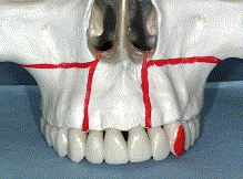
Fig. 1. Frontal view showing the lines of osteotomies on the maxillary complex.
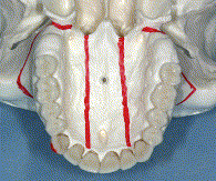
Fig. 2. Occlusal view of the maxillary complex showing the bilateral osteotomies
extending from the midpoint between the upper lateral and the canine (on the
right side) and between the upper central and lateral incisor (on the left
side). These osteotomies are extended to the posterior border of the maxillary
complex.
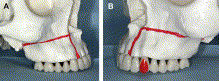
Fig. 3. Lateral views. A – right view, B – left view. The lateral osteotomy line
extends from the lower margin of the pirifrom aperture of the nasal cavity
towards the posterior margin of the maxillary complex.
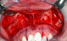
Fig. 4. This intra-oral photograph shows the OUF-RME lines of osteotomies in the
anterior region of the maxilla. It can be seen that two vertical osteotomies
extend from the horizontal osteotomies towards the interdental alveoli between
the central and lateral incisors bilaterally.